Sections
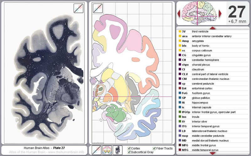
- High detailed anatomic coronal atlas of the human brain.
- (highlighted) anatomical structures
- The integrated nomenclature lets you explore the colored areas slice by slice.
- Any region of interest is also accessible with a search function either by name or abbreviation.
Access to the database and hierarchical tree will be available soon.
Virtual Microscopy: Cyto & Myelo Architecture

You may select the coronal sections by region or metric measures. Numbers indicate the position of the sections relative to the center anterior commissure.
With the option to navigate by anatomical names of all major structures and subdivisions of the brain or by using the "virtual microscope" you get access to high resolution sections.
Please notice that you need a Flash plugin to use the "virtual microscope".


Midsagittal view in Stereotaxic Space

Placement of the 69 sections depicted in the atlas. The intercommisural line (ICL) passes through the center of the anterior and posterior commissure, respectively. The vertical line (VCA) passes through the center of the anterior commissure.
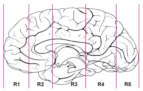
Lines indicate where the hemisphere was cut into 5 blocks (R1 - R5) before embedding.
Materials
The "Detailed Atlas of the Brain in Stereotaxic Space" is based on a brain from to the Vogt
collection in Düsseldorf. This brain was selected for presentation in this atlas because numerous
researchers have analyzed and reported on its structure in the last 60 years (see
here).
Embedding: The brain was cut by O. Vogt into blocks oriented vertically to the
intercommissural plane before its embedding in paraffin. The values of some linear measurements are
given here.
Sectioning: Serial frontal sections of 20 µm thickness were prepared.
Staining: Most sections were stained with either cresyl violet or hematoxylin. Some additional
sections remained unstained. Some of these unstained sections were used for immunohistochemistry
(e.g., substance P; see Mai et al., 1986).
Estimates of Volume Changes: Volume changes due to formalin fixation are negligible as the
brain volume at the fixation time corresponds to the values determined at autopsy (see Longerich,
1989). It is therefore reasonable that the dimensions of the formalin fixed brain represent the in
vivo situation. Volumetric changes due to the histological preparation (dehydration, paraffin
embedding, cutting, and mounting of the sections) were calculated from differences in linear
dimensions between the fixed, unembedded hemisphere (scaled photographs) and the serially sectioned
hemisphere. Lange and Thörner (1974) and Sievert (1992).
Photographic Plates and Corresponding Diagrams: The photographs and the diagrams always show
the entire hemisphere and the metric dimensions of the sections.
For the delineations in the diagrams we have always considered the results from the numerous cyto-
and myeloarchitectonic studies that were previously performed on the represented brain and we have
used as often as possible the original delineations provided by these earlier workers. Generally, the
consistency of the drawings, section-to-section, was of higher importance than a precise correspondence
between the photographs and the drawings.

